
Organellar Ribosomes
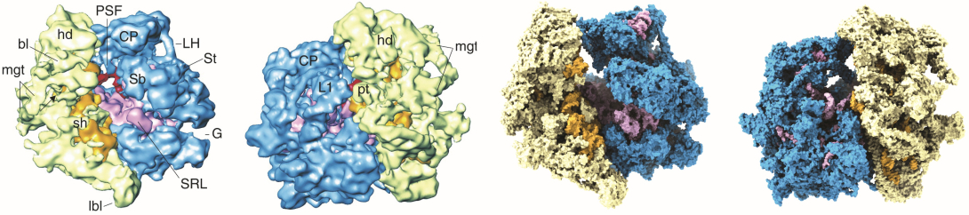
The Agrawal Lab was the first to determine the cryo-EM structure of an organellar ribosome. By determining the structure of a mammalian (Bos taurus) mitochondrial ribosome[1] (mitoribosome), his group discovered a dramatically altered overall architecture and distribution of its components such as ribosomal RNAs (rRNAs) and ribosomal proteins (r-proteins), as compared to bacterial ribosomes. Their structural analysis allowed them to make several discoveries, including:
- In sharp contrast to decades old belief, i.e., since the discovery of mitoribosome in mid 1960s, that the additional and enlarged mito-specific ribosomal proteins (MRPs) in the mammalian mitoribosome physically replace the drastically shortened mito-rRNAs, the Agrawal group discovered that most MRPs instead acquire novel quaternary positions giving the mitoribosome its unique shape that is relevant to mito-specific function.
- Two mitoribosomal subunits are held together by greater participation by MRPs, as compared to that in previously determined bacterial ribosomes. Also, the inter-subunit space, where mito-tRNAs and translational factors interact during protein synthesis, is significantly remodeled and dominated by MRPs, including the presence of a P-site finger.
- The presence of a gate-like MRP feature at the entrance of the mRNA channel in mammalian mitoribosome, possibly required for recruitment of leaderless mitochondrial mRNAs.
- That the lower two-thirds of the nascent-polypeptide exit tunnel (NPET) is dramatically remodeled, as compared to that in cytoplasmic ribosomes: NPET in mitoribosome is predominantly lined by MRPs, possibly to help create a unique conduit for hydrophobic nascent polypeptides and their co-translational insertion into the inner mitochondrial membrane.
These findings were confirmed in subsequent high-resolution cryo-EM studies of the mitoribosomes and their functional complexes from different mammalian species, including human mitochondria, by others and his own group[2], as the cryo-EM technology improved with the advent of the direct-electron detecting cameras.
By determining the cryo-EM structure of a chloroplast ribosome[3] (chlororibosome), the group revised the decades old assignment of a plastid-specific ribosomal protein 1 (PSRP1) to a chlororibosome-binding hibernation-promoting factor. [4]