
Derbyshire and Gray Laboratory Gallery
Video Gallery
ESX-1 primarily associates with the old “non-septal” pole. M. smegmatis cells expressing a fusion of msmeg0046::yfp were imaged every 10 mins in a heated growth chamber. The fluorescent fusion protein (red) localizes to a single cell pole, the old pole (Wirth et al., Mol. Micro. 2012).
Image Gallery
 [1]
[1]
Transconjugant colonies with different morphotypes result from a cross between M. smegmatis strains MKD24 and Jucho
 [2]
[2]
A transconjugant morphotype; colonies of a purified transconjugant isolated from a cross between M. smegmatis strains MKD24 and Jucho
 [3]
[3]
A transconjugant morphotype; colonies of a purified transconjugant isolated from a cross between M. smegmatis strains MKD24 and Jucho
 [4]
[4]
An M. smegmatis biofilm containing donor cells, mc2155
 [5]
[5]
A biofilm containing both donor and recipient cells of M. smegmatis
 [6]
[6]
A fluorescent image of the edge of an M. smegmatis colony expressing a mycobacterial gene fusion to the fluorescent protein dendra.
 [7]
[7]
A phase contrast image of the edge of an M. smegmatis colony expressing a mycobacterial gene fusion to the fluorescent protein dendra
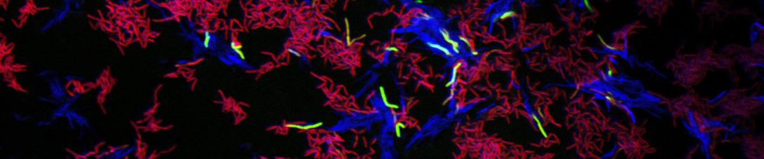
 and recipient (red) M. smegmatis cells grown in coculture. The recipient strain contacts a gfp reporter, which is activated (green) when donor and recipient cells are in direct cell contact.") [8]
[8]
Fluorescent image of a mixture of donor (blue) and recipient (red) M. smegmatis cells grown in coculture. The recipient strain contacts a gfp reporter, which is activated (green) when donor and recipient cells are in direct cell contact.
 and recipient (red) M. smegmatis cells grown in coculture. The recipient strain contacts a gfp reporter, which is activated (green) when donor and recipient cells are in direct cell contact.") [9]
[9]
Fluorescent image of a mixture of donor (blue) and recipient (red) M. smegmatis cells grown in coculture. The recipient strain contacts a gfp reporter, which is activated (green) when donor and recipient cells are in direct cell contact.